
Realtime MRI
Together with the Institute of Aerospace Medicine, the Institute for Software Technology is developing research software that enables the precise analysis of the cardiovascular system during free breathing. The methods developed for this purpose for the fully automatic evaluation of real-time MRI images are used both for the therapy and research of rare heart diseases on Earth, as well as for the research of effects and measures for long-term missions in space.
In cardiovascular medicine, the field of medicine that deals with diseases of the cardiovascular system, imaging techniques such as magnetic resonance imaging (MRI) are essential. In this process, large magnetic fields are used to resonantly excite certain atomic nuclei and thus produce sectional images of the human body. Today, new developments make it possible to record individual slices in a few tenths of a second as so-called real-time MRI, so that videos of the heart can be recorded under free breathing.
Realtime MRI images of a beating heart
Your consent to the storage of data ('cookies') is required for the playback of this video on Quickchannel.com. You can view and change your current data storage settings at any time under privacy.
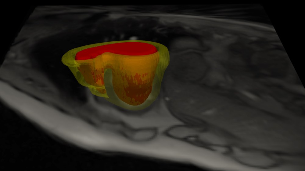
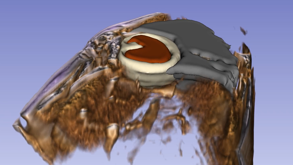
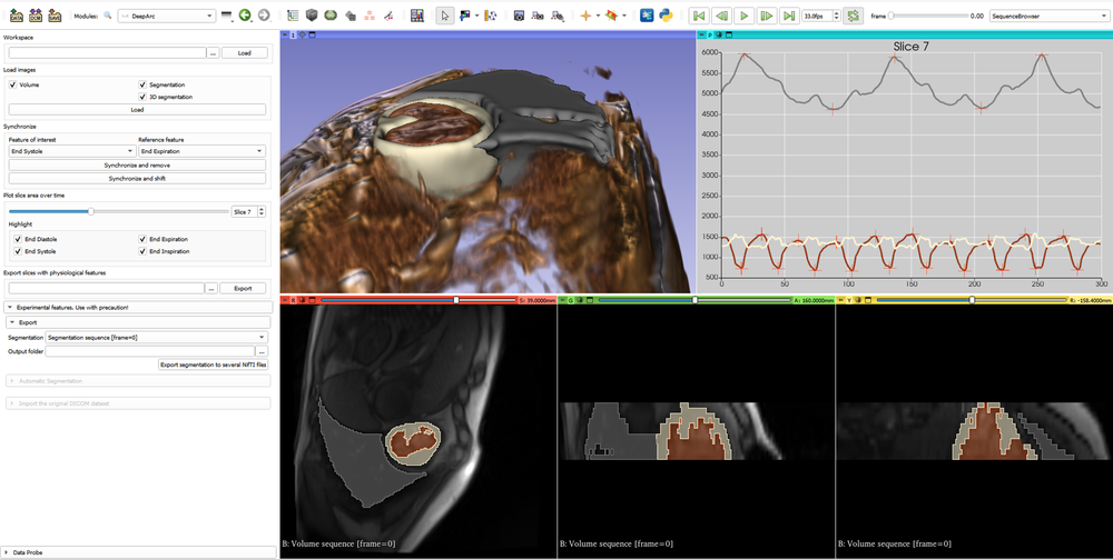
In the project, the researchers use modern AI algorithms to provide an automatic segmentation pipeline that can be used by research institutions and clinics to efficiently evaluate these videos. For this purpose, neural networks are trained for specific applications. In this way, pipelines are created that recognize the complex anatomies of severe congenital heart defects, or various influences of simulated weightlessness on the hearts of astronauts. These trained networks are used, for example, to find the blood volume in the ventricle and the volume of the surrounding heart muscle in the MRI images or videos. To do this, the AI classifies each pixel as either heart muscle, blood or background. Based on the predictions of these neural networks, functionalities are provided to evaluate pixel-by-pixel segmentation.
In this way, the program enables clinically relevant parameters, such as stroke volume, to be indicated in different respiratory phases. These parameters help both pediatric cardiologists to gain a deeper insight into the clinical picture and its development in general and for individual patients, as well as researchers at DLR. By developing a plug-in for the open source software "3DSlicer", the researchers can access many algorithms and functions already developed during software development.

Project runtime:
- 2021 - 2024
Scientific participants: